Research in Biology

Behavioral Response of Mud Crab Megalopae to Chemical Cues from Fish Species and Adult Conspecifics

By Jerelle Jesse


During the summer of 2014 OUR funded my honors research with Dr. Nancy O’Connor. This research has recently been published in the Journal of Experimental Marine Biology and Ecology and in what follows, I provide a summary of this research for the OUR blog readers.

Snapshot from the official website of the Journal of Experimental Marine Biology and Ecology, where Jesse’s research results were published

Portrait of Jerelle Jesse

§


In Massachusetts, Asian shore crabs have become more abundant than native mud crabs. Crab survival can be enhanced by antipredator behaviors in response to chemical cues released by predators.

Asian shore crab


The purpose of this study was to determine if and how mud crab megalopae (the last larval stage of the crab) respond to chemical cues from local fish predators and adult crabs of the same species and to understand the way local mud crab megalopae behaviorally respond to chemical cues. The study focused mainly on the importance of early life stages, the origin of the chemical cues, and their ability to respond to chemical stimuli. This could potentially shed light on how an invasive species can be more successful than a native species in this habitat.


Mud crab megalopa


Female egg-bearing mud crabs were collected from the rocky intertidal habitat during low tide periods. When the females became close to releasing larvae she was transferred to a small finger bowl, then placed in the incubator.

Egg-bearing female mud crab


Once the larvae were released they were cared for until they reached the megalopae stage when they were designated to an experiment.

Incubator with glass bowls of mud crab larvae before reaching the megalopal stage as well as females almost ready to release larvae in small glass finger bowls

§


Chemical cues for the experiment were made by the fish species or adult mud crabs being held in artificial seawater tanks to let their cue release into the water. The chemical cue seawater flowed through the apparatus, a glass pipe-shaped piece of equipment with an inflow opening, outflow opening, and a middle opening on top. The middle opening was to drop the individual megalopa into the apparatus with the cue flowing through.

The chemical cue flowed from the reservoir to a flow stabilizer, then a glass apparatus, and finally the sink. The megalopae were dropped into the middle funnel shaped opening in the apparatus


Once the megalopae was dropped into the apparatus it displayed 1 to 3 different behaviors then flowed out into the sink. The behaviors were categorized based on the orientation to the flow, the limb position, and the action performed. These behaviors included: control swim, random swim, perimeter swim, cyclone swim, closed roll, open roll, swim out, sideways walk run, slide, and push.

Left: Control swim; right: this megalopa happens to be on its back


The data were analyzed using generalized linear modeling. The results show no difference in behavioral responses between the two mud crab species. However, more open rolling behavior was seen for the mummichog cue, and significantly more walking on the bottom was seen for the adult cue. This indicates that megalopae can detect and respond to chemical cues in their environment. Megalopae can also tell the difference between adult conspecific cues and predator cues, and they can perform a different behavioral response depending on the cue.


§


My research experiences in Dr. Nancy O’Connor’s lab are some of my best memories from my time at UMass Dartmouth. I had so much fun conducting the research that summer, then rising to the challenge of analyzing the data, and ultimately getting the opportunity to present my work at multiple conferences. It was a rewarding experience that made my career at Umass Dartmouth truly special. Currently, I am working for the Division of Marine Fisheries and applying to graduate schools. I know that this research helped me become better prepared for fieldwork and graduate school. Being able to work with a master’s student, Ami Araujo, while I was an undergraduate gave me insight to the process and hard work involved with graduate school. Without OUR’s help I would not have been able to conduct this research, and help fulfill my dream of working as a marine biologist and going to graduate school.


Research in Chemistry

Developing Inexpensive Catalysts for Buchwald Carbon-Nitrogen Couplings


By Diego Javier-Jimenez

Portrait of Diego Javier-Jiminez


Catalyzed cross-coupling reactions using aryl halide reagents have found a prominent role in synthetic chemistry. The most notable are carbon-carbon coupling reactions, for which Heck, Negishi and Suzuki received the Nobel Prize in 2010. Similar carbon-nitrogen couplings, known as Buchwald-Hartwig aryl-amination reactions, have also found great utility, with applications in natural product synthesis, medicinal chemistry, organic materials chemistry, and catalysis. The catalysts in almost all cross-coupling reactions are based upon the precious metal palladium (price: $58,000/kg). Our lab is currently exploring different routes for the formation of carbon-nitrogen bonds with less expensive metals. This summer, I studied one such reaction in detail, analyzing the mechanism that the reaction follows.

§


My summer research involvement at UMass Dartmouth has been one of the most rewarding experiences of my undergraduate career. I had the pleasure of working with knowledgeable lab mates who were always willing to help, explain, and teach any skills necessary for me to be successful in my research. I would like greatly thank Dr. David Manke, working with him has inspired me to become the best chemist that I can and more. The experience has also significantly reaffirmed my goals of going to graduate school to obtain a Ph.D. in Chemistry. I would like to thank the Office of Undergraduate Research for funding this research, without their aid, this research experience would have not been possible. We are currently preparing two manuscripts that we hope to submit to peer-reviewed journals for publication this fall. I plan on continuing this work for the remaining two years at UMass Dartmouth, and hope that my research accomplishments will make me competitive for an NSF graduate research fellowship. The OUR has given me the opportunity to follow one of my life-long goals.

Research in Biology

Identification of SIAT7 in Symbiotic Clownfish and a Closely Related Non-Symbiotic Fish


By Deborah Dele-Oni

Portrait of Dele-Oni

Clownfish live in a close symbiotic relationship with sea anemones. This relationship is often used as a teaching tool for students to learn about ecology, evolutionary mutualism, and species interactions. This mutualistic relationship may be due to a sugar the anemones detect in the mucus of the prey species. An enzyme class known as sialyltransferases has been studied because of its importance of sea anemone recognition of prey. This class of sialyltransferases adds chains of sugars to proteins found in mucus. Clownfish may lack a specific type of sialytransferases known as SIAT7, which could be a factor as to why the clownfish do not get stung. However, although SIAT7 was not initially seen does not mean it is not there; rather it could indicate inactivation. Alternatively, clownfish may have SIAT7 in their genomes but may not express it in the cells that make the external mucus. To test this, I am studying both symbiotic and closely related non-symbiotic species to determine if SIAT7 is present in these species. I hypothesize that SIAT7 will be present in both the symbiotic clownfish and non-symbiotic closely related species but is inactive in the skin of symbiotic species. My goals were to test primers on tissues of anemonefish and closely related non-symbiotic species to see where expression occurred.


To accomplish these goals, I will:

  • Use degenerate PCR to obtain partial sequences of SIAT7 from symbiotic and non-symbiotic fish species.
  • Use inverse PCR to determine the sequences of the regions surrounding the SIAT7 gene.
  • Use quantitative PCR to determine which tissues express SIAT7 in symbiotic and non-symbiotic fish species.


To approach this, I knew that SIAT7 had been identified in close relatives of clownfish. If primers were designed based from those sequences and added to DNA of symbiotic clownfish, there would be a product formed if the primers found complementary parts of the DNA. In the spring, I completed degenerate PCR to try and acquire partial sequences of SIAT7 from non-symbiotic fish species. The degenerative primers were created from the bicolor damselfish (Stegastes partitus; Genbank accession XP_008298796.1), and PCR was done on cDNA samples from the ocellaris clownfish (Amphiprion ocellaris) and the non-symbiotic Springer’s damselfish (Chrysiptera springeri) which is a close relative. The PCR yielded some products which are bright bands in the gel below (Figure 1). The brighter the bands the more concentration of DNA, showing successful replication. These samples were then cleaned up and sent off for sequencing. The sequencing results were crosschecked with the NCBI database and matches that appeared were not of SIAT7.  Instead they matched to other genes like protein FAM20A isoform X3 inform the Southern pig-tailed macaque or monkey (Macaca nemestrina) (Figure 2) or to bacterial genes like protein A2680_02525 from the bacteria, Candidatus kaiserbacteria . These sequencing results are the DNA of one of the bands from the failed attempt using degenerate PCR. These results indicate that our DNA in the degenerate PCR was not successful at producing a partial part of the SIAT7 gene.

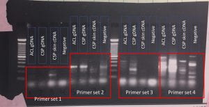
Figure 1. Gel showing the results of degenerate PCR searching for SIAT7 in springers damselfish (chrisyptera springeri, CSP) and clarks clownfish (amphiprion clarkia, ACL) genomic (gDNA) and skin cDNA samples.


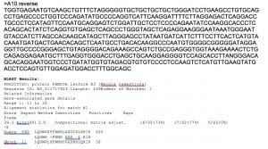
Figure 2. Sequencing results and BLASTx alignment for a sample.  The BLAST results show a match with the protein FAM20A isoform X3 with the Southern pig-tailed macque (Macaca nemestrina) which is a medium-sized monkey.


Since the degenerate PCR primer was not successful at yielding a partial sequence for SIAT7, another approach to obtaining this sequence was taken. Marian Wahl, a graduate student in Dr. Robert Drew’s lab, had recently sequenced transcriptomes from several species of anemonefish and non-symbiotic fish.  Transcriptomes are all of the RNA that is made by genes of an organism. This is of interest because it shows exactly what is made and what will potentially be translated to proteins. This was not available in the spring but became available early this summer. I redesigned primers for four species of anemonefish (list species) and non-symbiotic fish (list species) to be used in the PCR. This provided me with a better chance of getting PCR product because the primers used in the PCR were designed from the exact species they would be testing in. Also, I would be able to see right away if SIAT7 was really present in the fish species because I would be checking their transcriptomes to see if it was present or not. If SIAT7 was present, I would get a gene sequence from the transcriptomes.


To do this a reference gene was identified from the bicolor damselfish (Stegastes partitus; XP_008298796.1). This reference gene was used to find matching sequences from the transcriptomes of the study species using Local BLAST. I found that SIAT7 appeared in all species transcriptomes that were checked. From this, I could say that SIAT7 is found in both symbiotic and non-symbiotic species of fish. However, the specific tissue or tissues it is expressed in and to what extent was not known from this information.


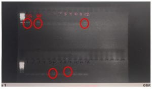
Figure 3. PCR Results using primers designed for Amphirion clarkii (ACL) species and Amblyglyphidodon curacao (AmCu) species.


After going back to look at the specific gene sequences that were used to make the primers, there was evidence that SIAT7 across these species may be paralogs. Paralogs are genes that have evolved by duplication events, resulting in two copies of the gene in different locations of the genome.  After duplication, these copies evolve independently, accumulating different mutations. After a long period, these paralogs may still encode for the same protein but can have very different DNA sequences. This is interesting to note because it could be evidence that clownfish symbiosis caused this duplication event to occur. We found paralogs in the Clarks clownfish but not in the other three species we tested which were the staghorn damselfish (Amblyglyphidodon Curacao), the three spot damselfish (Dascyllus Trimaculatus) and the three stripe damselfish (Dascyllus Aruanus). We found this out by aligning the different transcript that were gotten from the Local BLAST. When aligned I found that the Clarks clownfish transcripts with similar trinity numbers (numbers that appear after the letters “DN” in Figure 4) were more closely related than the ones with dissimilar numbers. If the sequence used to make the primers were made using one paralog, other paralogs will not be accounted for in the study and the PCR will not yield consistent results.


To account for paralogs, some bioinformatics was done to identify exactly where duplication events might have occurred and in what species. To do this, transcriptomes for the species of interest were identified and aligned to each other using computer programs such as MUSCLE, TranslatorX, and the NCBI Blast Website (Figure 4). When transcriptomes are aligned, the programs will put similar sequences together and dissimilar sequences further apart from each other.  This figure highlights that species with the same sequences (samples with the same Trinity numbers) may be from the same gene. For example, the sequences DN83440 and DN182523 from A. clarkii are probably paralogs but there are two copies of DN182523 which are probably splice variants or have alternative transcription start sites.


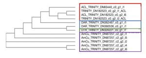
  Figure 4. Phylogenetic tree of SIAT7 cDNA samples from Clark’s Clownfish (ACL), Three stripe domino damselfish (DAR), Three spot domino damselfish (DTR) and staghorn damselfish (AmCu).


There is still much to do so I am continuing work on this project this fall.  This figure will be updated to include some well-studied fish and re-rooted to provide more accurate results. Some cichlid fish are more well understood in the evolution of fish, and using these as references for our SIAT7 sequences, can provide me with some information on paralogs. Once paralogs are completely identified, more specific primers can be designed that will hopefully yield consistent PCR results. Another approach that will be taken is to align protein sequences. Right now, the aligning that has been done has used cDNA sequences.


§


From the work done this summer, I can say that SIAT7 is found in symbiotic and non-symbiotic fish that I studied, indicating that clownfish did not lose SIAT7 as part of the evolution of symbiosis with sea anemones.  However, I detected evidence of gene duplication which introduced paralogs. Going forward, I seek to understand when these duplication events occurred and if it is related to the clownfish-sea anemone symbiosis. I am looking to seeing if the evolution of paralogs in SIAT7 allowed anemonefish to live symbiotically with anemones or if it is completely unrelated to this.

§


This summer research experience has provided me with an opportunity to continue with a long-term research experience. I stepped out of my comfort zone and experienced new things in the lab and learned immensely from bioinformatics alone. Being able to get results from looking at gene sequences and databases on local computers, and searching national gene databases, I could answer one of my research questions without even picking up a pipette. As a biology student, I underestimated the wealth of information bioinformatics shows and how important it is to do these steps in research.  Conducting experiments in the lab is rewarding but interpreting the data, and understanding it is the main goal. This summer research experience, I learned to think about long term goals and the bigger picture. Having participated in only short-term research experiences before, I was usually just thrown into a situation where I had to think quickly on my feet and do a series of experiments and interpret my immediate results. However, being at UMass during the summer, I could continue work I had started before. This allowed me to see what a long-term project entails. Data interpretation and relating results to a goal is something that I have strongly developed this summer. I feel much more prepared to pursue more long-term projects. I have developed myself as a critical thinker and a troubleshooter in my research and found a new appreciation for the study of bioinformatics.


Research in Marine Biology

Influence of UV Light on Marine Biofilms

By Alexandria E. Profetto


Currently I am a rising junior marine biology major at UMass Dartmouth. My career here at the university started late due to being a member of the Massachusetts Army National Guard. After delays from training and a deployment from 2014-2015, I could begin my long sought after pursuit of a degree in marine biology. Thanks to the funding from the OUR and additional assistance by the Dean’s Undergraduate Fellowship, I have been able to work on an antifouling project, originally started in 2016 by Boston Engineering Corporation (BEC) and Dr. Pia Moisander at the Biology Department. The project was focused around the reduction of growth on marine biofilms, specifically on capabilities of a prototype device, developed by BEC, based on LED-generated ultraviolet (UV) light for use as an antifouling method for ship hulls (UV-C band light).

Portrait of Alexandria E. Profetto (left) as a member of the National Guard


Biofilms can be found and formed on a variety of surfaces, varying from indwelling medical devices to natural aquatic systems. Formation of a biofilm (“fouling”) begins with an accumulation of microbial cells on a surface surrounded in a polysaccharide based matrix. Depending on the environment in which the biofilm has formed, non-cellular materials such as clay or silt particles can be found in the matrix (Donlan, 2002). In aquatic based biofilms, the solid-liquid boundary between water and the surface, such as a ship hull, offers an ideal environment for the attachment and growth of microorganisms. Bacteria and diatoms are the most dominate forms reported in biofilms and are coined as “microfoulers”. These microfoulers play a very important role by providing signals for the attachment of various macrofouling organisms ranging from algae and barnacles to oysters and polychaetes (Donlan, 2002). This can be a nuisance for aquaculturists as well as commercial and recreational fishermen. Traditionally, antifouling heavily relied on fouling-reducing marine paints that although reduced in toxicity, still contain some toxic chemicals which can potentially cause harmful environmental impacts. Limited options for environmentally friendly and effective eradication of biofilms have created a need for alternative antifouling methods (Kim et. al, 2016).


Left: 30C Plates post sampling one week into the experiment; right: 30C Experiment Bin


During my project over the summer of 2016, we had a few goals regarding methodology, toward development of a repeatable and controlled experimental system for growing marine biofilms in the lab. We also wanted to test the capabilities of the UV device on biofilms grown under a range of temperatures, using microalgal cultures isolated from Buzzards Bay by Dr. Moisander in 2016. The biofilms were grown for 1-2 weeks in 32L of inoculated microalgal cultures at two temperatures. Forty aluminum plates, painted to simulate a boat hull, with non-antifouling paint, were used to grow the biofilms on. At specified times, the plates were treated with the UV light with one of the three duration times (1, 10 or 20 minutes) and then placed back in the bin to continue growth. Triplicate plates were included for each treatment.  Samples were then collected from the treated and non-treated areas (one and two weeks after the UV treatment) to be analyzed at a later date. Samples were collected to investigate presence of chlorophyll a (representing microalgal abundance) and abundances of bacteria on the surfaces. A second experiment was conducted with bacterial mixed cultures in one temperature only and a 1-week post-treatment incubation.

§


 Left: Front View of Setup with 22C Bin; right: Plate Arrangement of 22C Bin


By the end of the summer, all samples were collected for each analysis and experiments completed. I also finished the analysis of all chlorophyll samples using fluorometry, and started the bacterial counts using epifluorescence microscope. The data compilation for chlorophyll data is currently in progress, and I am continuing to complete the bacterial abundance counts over the next few months.


Left: 22C Plates after 1 week after the first UV treatment; right: 3D printed plate holder (by Boston Engineering) used for precisely sampling the plates


Overall, the UV device appeared to be successful in killing existing biofilm and slowing down regrowth in the already formed biofilms. The observations show that we were successful in creating artificial marine biofilms in the lab and demonstrate the effectiveness of the UV device on these biofilms, mirroring overall results from pilot experiments conducted by Moisander lab and the BEC collaborators with natural biofilms from Buzzards Bay in 2016.

UV device setup on top of plate prior to treatment

§


My research experience this summer was very eye opening regarding where and how I want to work in my future research career. I thoroughly enjoyed coming up with an experimental design and tackling the research challenges with Dr. Pia Moisander, as well as seeing the project come to a successful completion. Without her mentoring filled with her wealth of knowledge and expertise, I can’t say my problem solving and critical thinking in terms of science would have progressed as well as I’ve noticed. Collaborating with other members of the lab team with in person lab meetings were truly priceless experiences that I am so grateful for being afforded. Getting other opinions, ranging from an REU undergraduate to a post doc, was a great way to expand my thinking on my project than to just “what does is this data?”. My hope for this upcoming academic year is to continue assisting with this biofilm project or any project, finishing up data analysis and learn as much as I can from Dr. Moisander and her three Ph.D. students. I’d also like to thank visiting post-doc Mar Benavides and REU undergraduate Clay Evans for allowing me to bounce ideas off them as well as learn from their research projects.


References

Donlan, Rodney M. “Biofilms: Microbial Life on Surfaces.” Emerging Infectious Diseases 8.9 (2002): 881-90. Web.

Kim, Minhui, Shin Young Park, and Sang-Do Ha. “Synergistic Effect of a Combination of Ultraviolet–C Irradiation and Sodium Hypochlorite to Reduce Listeria Monocytogenes Biofilms on Stainless Steel and Eggshell Surfaces.” Food Control. Elsevier, 03 May 2016. Web.