

Research in Marine Biology
Influence of UV Light on Marine Biofilms
By Alexandria E. Profetto
Currently I am a rising junior marine biology major at UMass Dartmouth. My career here at the university started late due to being a member of the Massachusetts Army National Guard. After delays from training and a deployment from 2014-2015, I could begin my long sought after pursuit of a degree in marine biology. Thanks to the funding from the OUR and additional assistance by the Dean’s Undergraduate Fellowship, I have been able to work on an antifouling project, originally started in 2016 by Boston Engineering Corporation (BEC) and Dr. Pia Moisander at the Biology Department. The project was focused around the reduction of growth on marine biofilms, specifically on capabilities of a prototype device, developed by BEC, based on LED-generated ultraviolet (UV) light for use as an antifouling method for ship hulls (UV-C band light).
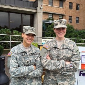
Portrait of Alexandria E. Profetto (left) as a member of the National Guard
Biofilms can be found and formed on a variety of surfaces, varying from indwelling medical devices to natural aquatic systems. Formation of a biofilm (“fouling”) begins with an accumulation of microbial cells on a surface surrounded in a polysaccharide based matrix. Depending on the environment in which the biofilm has formed, non-cellular materials such as clay or silt particles can be found in the matrix (Donlan, 2002). In aquatic based biofilms, the solid-liquid boundary between water and the surface, such as a ship hull, offers an ideal environment for the attachment and growth of microorganisms. Bacteria and diatoms are the most dominate forms reported in biofilms and are coined as “microfoulers”. These microfoulers play a very important role by providing signals for the attachment of various macrofouling organisms ranging from algae and barnacles to oysters and polychaetes (Donlan, 2002). This can be a nuisance for aquaculturists as well as commercial and recreational fishermen. Traditionally, antifouling heavily relied on fouling-reducing marine paints that although reduced in toxicity, still contain some toxic chemicals which can potentially cause harmful environmental impacts. Limited options for environmentally friendly and effective eradication of biofilms have created a need for alternative antifouling methods (Kim et. al, 2016).
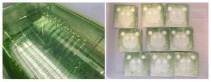 Left: 30C Plates post sampling one week into the experiment; right: 30C Experiment Bin
During my project over the summer of 2016, we had a few goals regarding methodology, toward development of a repeatable and controlled experimental system for growing marine biofilms in the lab. We also wanted to test the capabilities of the UV device on biofilms grown under a range of temperatures, using microalgal cultures isolated from Buzzards Bay by Dr. Moisander in 2016. The biofilms were grown for 1-2 weeks in 32L of inoculated microalgal cultures at two temperatures. Forty aluminum plates, painted to simulate a boat hull, with non-antifouling paint, were used to grow the biofilms on. At specified times, the plates were treated with the UV light with one of the three duration times (1, 10 or 20 minutes) and then placed back in the bin to continue growth. Triplicate plates were included for each treatment. Samples were then collected from the treated and non-treated areas (one and two weeks after the UV treatment) to be analyzed at a later date. Samples were collected to investigate presence of chlorophyll a (representing microalgal abundance) and abundances of bacteria on the surfaces. A second experiment was conducted with bacterial mixed cultures in one temperature only and a 1-week post-treatment incubation.
§
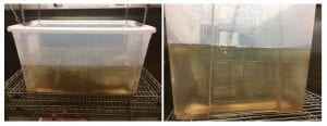
Left: Front View of Setup with 22C Bin; right: Plate Arrangement of 22C Bin
By the end of the summer, all samples were collected for each analysis and experiments completed. I also finished the analysis of all chlorophyll samples using fluorometry, and started the bacterial counts using epifluorescence microscope. The data compilation for chlorophyll data is currently in progress, and I am continuing to complete the bacterial abundance counts over the next few months.
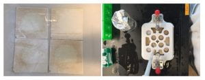 Left: 22C Plates after 1 week after the first UV treatment; right: 3D printed plate holder (by Boston Engineering) used for precisely sampling the plates
Overall, the UV device appeared to be successful in killing existing biofilm and slowing down regrowth in the already formed biofilms. The observations show that we were successful in creating artificial marine biofilms in the lab and demonstrate the effectiveness of the UV device on these biofilms, mirroring overall results from pilot experiments conducted by Moisander lab and the BEC collaborators with natural biofilms from Buzzards Bay in 2016.
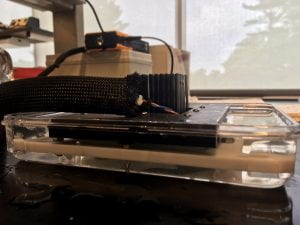
UV device setup on top of plate prior to treatment
§
My research experience this summer was very eye opening regarding where and how I want to work in my future research career. I thoroughly enjoyed coming up with an experimental design and tackling the research challenges with Dr. Pia Moisander, as well as seeing the project come to a successful completion. Without her mentoring filled with her wealth of knowledge and expertise, I can’t say my problem solving and critical thinking in terms of science would have progressed as well as I’ve noticed. Collaborating with other members of the lab team with in person lab meetings were truly priceless experiences that I am so grateful for being afforded. Getting other opinions, ranging from an REU undergraduate to a post doc, was a great way to expand my thinking on my project than to just “what does is this data?”. My hope for this upcoming academic year is to continue assisting with this biofilm project or any project, finishing up data analysis and learn as much as I can from Dr. Moisander and her three Ph.D. students. I’d also like to thank visiting post-doc Mar Benavides and REU undergraduate Clay Evans for allowing me to bounce ideas off them as well as learn from their research projects.
References
Donlan, Rodney M. “Biofilms: Microbial Life on Surfaces.” Emerging Infectious Diseases 8.9 (2002): 881-90. Web.
Kim, Minhui, Shin Young Park, and Sang-Do Ha. “Synergistic Effect of a Combination of Ultraviolet–C Irradiation and Sodium Hypochlorite to Reduce Listeria Monocytogenes Biofilms on Stainless Steel and Eggshell Surfaces.” Food Control. Elsevier, 03 May 2016. Web.