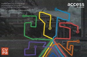
Research in Computer & Information Science

Recovery of Fine Details for Fast Imaging Knee Pathologies

By Jasina Yu

Portrait of Jasina Yu


Knee diseases or injuries are very common in the United States. For example, more than 14 million Americans suffer from knee osteoarthritis. Magnetic resonance imaging (MRI), as an interdisciplinary field of computer science, mathematics, engineering, and MR physics, provides an accurate noninvasive assessment of knee pathology. The soft tissue structures (such as menisci, ligaments, and cartilage) and bone marrow of the knee can be visualized for diagnosis and prognosis. However, an MRI scan generally needs 45-90 minutes. As a freshman, I am interested in computer science, mathematics, and physics. MRI integrates those fields, and it is a great research topic to achieve my study goals.

The objective of the project is to advance our understanding of MRI by keeping fine details of knee images. Knee pathologies are accurately visualized without sacrificing imaging speed. The fundamental understanding of the feature representation, extraction, and selection in the artificial intelligence (AI)-based reconstruction process will benefit the knee pathology features’ recovery from highly undersampled data. Detailed information lost in the reconstruction process was studied. This project has helped initiate my research activities at UMD and I hope to advance my career as a researcher and innovator in biomedical imaging investigation.


Based on the preliminary research using the fastMRI dataset [1], our AI-based technique (as shown in the 4th column of the figure above) can recover more details than the other two methods shown in the 2nd and 3rd columns of the figure. The reference knee image is shown in the 1st column of the figure. Our AI method has closer pathological details to the reference knee image because the other two methods have degraded image quality.


Reference

[1]. Knoll F, et. al. fastMRI: a publicly available raw k-space and DICOM dataset of knee images for accelerated MR image reconstruction using machine learning. Radiol Artif Intell. 2020;2(1): e190007.

Research in Bioengineering

Cell Viability of Novel Wound Healing Hydrogels

By Abid Neron


Introduction

Over the summer and my fall semester, I was culturing cells. I started off completely clueless but slowly I started learning and gaining a better understanding of the ins and outs of cell culture. I should start off by explaining what cell culture actually is. Basically, it’s growing a certain cell outside of its normal environment, in a lab. Cell culture is used to study these cells’ growth patterns and how to change their rate of growth. There are multiple cell lines/types, such as skin cells, kidney cells, cancer cells, and much more. I was mostly culturing HEK293 cells which are mammalian kidney cells. I also cultured A375 cells as well, these are a skin cancer cell line. I started off doing cell culture by just following a list of steps telling me what to do. I didn’t understand what I was doing, though. After some research and passaging my cells multiple times, I started to get an understanding of what cell culture is and the steps started making sense. Finally, after perfecting my technique, I wanted to start research using them. I talked to Dr. Tracie Ferreira, and she tasked me with creating a protocol to test cell growth on certain substances. This protocol would be used by the seniors on their final projects. I was introduced to the seniors and their projects and collaborated with them using my knowledge of cell culture.

Abid Neron passaging his cells inside a biosafety cabinet


Methods

Creating a standard protocol to test cell growth (Also known as cell viability) took a lot of research and multiple failed experiments but with each failed experiment I went back to the drawing board and tweaked some steps until I finally found the best method to test cell viability.

My cell viability test uses a substance called Resazurin which is a special dye that changes color depending on cell activity. Cells produce ATP. Resazurin changes color based on the amount of ATP produced. As cell number increases, so does the production of ATP. Further, Resazurin doesn’t affect cells’ growth and doesn’t damage them unlike other cell viability tests. Using a Spectrometer, I can analyze the change in color for each test. The larger the value the more cells were growing. After testing my protocol on students’ substances called Hydrogels, the test offered good results and could be replicated multiple times to get more accurate results if needed. Seniors are currently using the protocol I created to test cell viability on their Hydrogels.

I had an issue with bacteria growing on the hydrogels which would mess up the test, so after multiple experiments, I found the best way to sterilize the hydrogels and keep bacteria away from the cells by using different plates everyday to get more accurate results.

Cell Viability Test

(Notice how the bottom right well is a brighter color, that means that the cells are growing faster!)


Results

I tested my protocol on multiple hydrogels and compared their growth with cells growing under optimal conditions:

A hydrogel’s cell viability over 5 days


Discussion

My research is still ongoing and I’m constantly furthering my knowledge of cell culture and perfecting my technique. Cell culturing has become an almost therapeutic process for me. While there’s a lot to learn, it’s always nice to apply what I’m learning about. Researching cell culture is an incredible experience that I truly loved and sharing my experience with other students and teaching them cell culture and seeing their awe when they see cells growing is always rewarding. I wouldn’t have had this experience if it weren’t for Dr. Trace Ferreira. She taught me everything I know about cell culture and is always there to bounce ideas with. Going forward, I will test more hydrogels and hopefully teach more students how to apply my cell viability test in their own research.

Research in Graphic Design & History of Architecture

Access@UMassD

by Eden McKenna-Bateman and Evan Grant

Exploring accessibility on campus was an interdisciplinary independent study, funded by the OUR. The project was focused on the architectural and spatial history of UMass Dartmouth and how *access* to spaces and resources has become available, modified, and contested– spatially and architecturally speaking– over the years. We first began by visiting the archives of the university, where important drawings and blueprints by its main architect Paul Rudolph (1918-1997) are housed. We wanted to know how Rudolph crafted his vision and in what ways he conceptualized accessibility. We then studied the physical transformation of the campus over the years and finally placed our focus on the current campus with added buildings and renovations that have modified the original design. In particular, we studied how physical access impacts the  function of our present day campus and its diverse communities. After conducting our architectural history research, we began to use our knowledge of graphic design to give an “image” to our research. The purpose of creating a bold image was to bring awareness about issues of accessibility, not only for special needs student population, but also for those students who wish to have better access to resources available on campus. Instead of doing our work behind closed doors, we wanted to engage students and faculty in our research. For the graphic design portion of this project we also explored the different ways in which we can make a more impactful presentation. We explored several activist projects on US campuses. We studied patterns, fonts, colors, and compositions that are employed in effective activist projects.


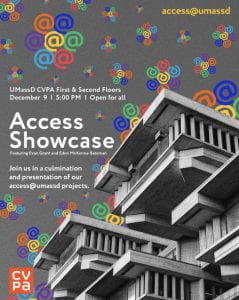
Poster designed by David Grant & Eden McKenna-Bateman for the final phase of their project.

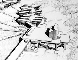
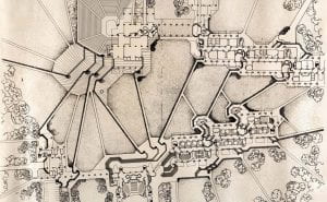
Paul Rudolph. Aerial views of the campus at the Southeastern Massachusetts Technological Institute (SMTI/UMass Dartmouth), ca. 1963. Ink on linen. Courtesy of UMass Dartmouth, Claire T. Carney Library, Archives, and Special Collections.


Our overall objective with this study was to create collaborative and interactive projects between students and professors addressing various accessibility issues on campus. The goal was to bring awareness to issues of accessibility and its significance for student success. A substantial part of this interactive project drew on the student body’s thoughts and opinions. While the campus meets ADA requirements, those requirements only fulfill the bare minimum due to many ADA regulations being outdated, and often do not fully meet the needs of students with disabilities. The inequality in accessibility at UMassD is non-inclusive. Our study was research-focused and project-based. Throughout the course of this project, we researched and conducted interviews/surveys, and collaborated with students and faculty of various professional fields to create and deploy four projects throughout our campus that address accessibility issues while allowing students to share their thoughts and opinions on the matter.


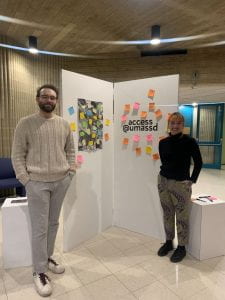
 Eden Mackenna-Bateman and Evan Grant and snapshots from their installations


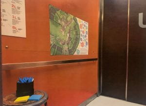
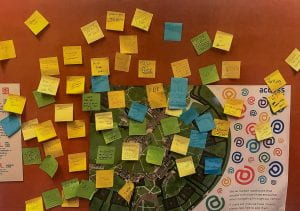
We secured funds from the OUR for multiple installation projects. We had a CVPA Elevator Survey, consisted of art installations in one elevator in each college as well as the library. The installations consisted of printed maps with roadblocks labeled, with cut vinyl applied within the elevator asking a question that students can respond to via sticky notes. In hopes of creating a broader scope to reach people, we promoted our project through pencils and stickers that spread awareness within the student body. Another installation project was Access Tours, consisted of an interactive experience that followed our original map of campus inaccessibility through the use of signage and pathway markers that allowed students and faculty to experience the roadblocks that people with disabilities face when navigating through our campus. Our third project was a collaboration between Access@UMassD and the University to integrate our findings into the interactive virtual campus map. Our fourth and final project, the Access@UMassD Exhibition presented our thesis through a display of all our research and documentation of our projects within a series of popup installations. This included the results of our first three projects, documenting student and faculty experiences, which consisted of stories, photographs and physical pieces from the installations, accompanied by a presentation and takeaway items such as information cards and pamphlets. We plan to present the outcome of this research to campus administration. Regardless of the final outcome, we want to take this opportunity to say that this research has been very informative. We learned how to conduct collaborative research projects; in our studies, we also learned about the ways in which architecture can limit or extend our access to resources and ideas; additionally, we learned how graphic design can play a significant role in various civic campaigns. We are grateful to our mentors Professor Pamela Karimi (Art History) and Michelle Bowers (Graphic Design) who guided us through this project. We are also grateful to the OUR that funded our project.