

Research in Computer & Information Science
Recovery of Fine Details for Fast Imaging Knee Pathologies
By Jasina Yu

Portrait of Jasina Yu
Knee diseases or injuries are very common in the United States. For example, more than 14 million Americans suffer from knee osteoarthritis. Magnetic resonance imaging (MRI), as an interdisciplinary field of computer science, mathematics, engineering, and MR physics, provides an accurate noninvasive assessment of knee pathology. The soft tissue structures (such as menisci, ligaments, and cartilage) and bone marrow of the knee can be visualized for diagnosis and prognosis. However, an MRI scan generally needs 45-90 minutes. As a freshman, I am interested in computer science, mathematics, and physics. MRI integrates those fields, and it is a great research topic to achieve my study goals.
The objective of the project is to advance our understanding of MRI by keeping fine details of knee images. Knee pathologies are accurately visualized without sacrificing imaging speed. The fundamental understanding of the feature representation, extraction, and selection in the artificial intelligence (AI)-based reconstruction process will benefit the knee pathology features’ recovery from highly undersampled data. Detailed information lost in the reconstruction process was studied. This project has helped initiate my research activities at UMD and I hope to advance my career as a researcher and innovator in biomedical imaging investigation.
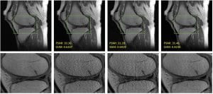
Based on the preliminary research using the fastMRI dataset [1], our AI-based technique (as shown in the 4th column of the figure above) can recover more details than the other two methods shown in the 2nd and 3rd columns of the figure. The reference knee image is shown in the 1st column of the figure. Our AI method has closer pathological details to the reference knee image because the other two methods have degraded image quality.
Reference
[1]. Knoll F, et. al. fastMRI: a publicly available raw k-space and DICOM dataset of knee images for accelerated MR image reconstruction using machine learning. Radiol Artif Intell. 2020;2(1): e190007.